Arthritis of the big toe — known medically as hallux rigidus — is the most common arthritic condition of the foot. It affects approximately 1 in 40 adults over the age of 50, and it can be profoundly disabling. The first metatarsophalangeal (MTP) joint carries roughly 119 percent of body weight with every step you take. When that joint is stiff, painful, and inflamed, the simple act of walking becomes a problem. Pushing off during gait hurts. Shoes become difficult to tolerate. Running, hiking, yoga, and countless other activities become impossible or miserable.
When conservative treatments — stiff-soled shoes, orthotics, anti-inflammatory medications — are no longer providing adequate relief, surgery becomes the next conversation. And for most patients, the first question is the same one I hear every day: How big is the incision? How much will it hurt? When can I walk?
Minimally invasive surgery is changing the answers to all three of those questions. Whether the goal is preserving the joint through a cheilectomy with Moberg osteotomy, or definitively eliminating pain through a first MTP fusion, percutaneous and minimally invasive techniques allow me to achieve the same — or better — clinical outcomes as traditional open surgery, with smaller incisions, less soft-tissue disruption, lower infection rates, less postoperative pain, and a faster return to walking.
Understanding Big Toe Arthritis: What Happens Inside the Joint
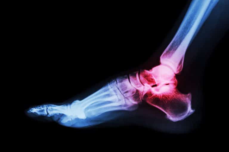
Hallux rigidus is a degenerative condition of the first MTP joint — the large joint at the base of your big toe where the first metatarsal bone meets the proximal phalanx. Over time, the smooth articular cartilage that lines the joint surfaces wears away, and bone spurs (osteophytes) form, particularly along the top (dorsal) surface of the joint. These bone spurs block upward motion of the toe, which is why the hallmark symptom is pain and stiffness when you push off during walking.
The condition typically develops between the ages of 30 and 60 and tends to affect women more than men. In most cases the cause is idiopathic — the result of genetics, foot structure, and cumulative mechanical stress rather than a single traumatic event. Nearly two-thirds of patients have a family history of the condition, and up to 79 percent have bilateral involvement. As the arthritis progresses, pain moves from being limited to the end-range of motion to becoming present through the middle arc of motion — a critical distinction that determines which surgical approach is most appropriate.
Hallux rigidus is graded on a scale from 0 to 4 (Coughlin and Shurnas classification), with mild grades (1 and 2) characterized by dorsal bone spurs with relatively preserved joint space, and advanced grades (3 and 4) showing significant cartilage loss, narrowed or obliterated joint space, and pain through midrange motion. This staging directly informs the surgical decision-making: milder disease is amenable to joint-preserving procedures, while advanced disease typically requires fusion.
Two Surgical Paths: Joint Preservation and Joint Fusion
For patients with mild to moderate hallux rigidus — those whose pain occurs primarily at the extremes of motion and whose joint space remains relatively preserved — the goal of surgery is to preserve the joint, eliminate the impingement, and restore comfortable range of motion. The two most effective procedures in this category are cheilectomy and cheilectomy with Moberg osteotomy.
For patients with advanced hallux rigidus — significant cartilage loss, midrange pain, and a joint that is grinding bone on bone — the gold standard remains arthrodesis (fusion) of the first MTP joint. Fusion eliminates the arthritic joint entirely by allowing the metatarsal and proximal phalanx to grow together into a single, stable, pain-free unit.
Both of these surgical paths have traditionally been performed through open approaches — long incisions, direct joint exposure, and significant soft-tissue dissection. Both can now be performed through minimally invasive and percutaneous techniques, and the advantages are substantial.
Minimally Invasive Cheilectomy and Moberg Osteotomy: Preserving the Joint Through a Keyhole
A cheilectomy is the removal of the dorsal bone spur (osteophyte) and approximately 25 to 30 percent of the dorsal metatarsal head — the portion of bone that is blocking upward motion of the toe and causing impingement. It is a straightforward concept: remove the mechanical block, and the joint moves freely again.
The Moberg osteotomy is a powerful complement to cheilectomy. It involves making a small dorsal closing-wedge cut in the proximal phalanx (the bone of the big toe itself), which tilts the toe slightly upward. This has two effects: it shifts the functional arc of motion into a more useful range for walking and push-off, and it moves the contact point of the joint away from the area of greatest cartilage damage. The combination of cheilectomy plus Moberg osteotomy has been shown to produce significant improvements in pain scores, functional outcomes, and dorsiflexion range of motion.
Traditionally, both of these procedures require a medial or dorsomedial incision of 4 to 6 centimeters, a formal capsulotomy (opening the joint capsule), and direct visualization of the bone spur and joint surface. This open approach provides excellent visualization but comes at the cost of soft-tissue disruption, postoperative swelling, and a recovery period that reflects the extent of the surgical dissection.
The percutaneous approach is fundamentally different. Through a small keyhole incision — typically 2 to 3 millimeters — a specialized surgical burr is introduced under fluoroscopic guidance. The dorsal osteophyte is resected percutaneously, and the Moberg osteotomy is performed through an additional small incision using a percutaneous saw, without opening the joint capsule or disrupting the surrounding soft tissues. The entire procedure is guided by live X-ray imaging, ensuring precise bone removal and accurate osteotomy placement.
The published evidence on percutaneous cheilectomy is encouraging. A 2024 systematic review of minimally invasive cheilectomy techniques, encompassing nearly 300 patients, found that dorsiflexion improved on average from approximately 32 degrees to over 61 degrees, with an overall complication rate of only 6.1 percent and a wound infection rate of approximately 2 percent. Patient satisfaction and functional outcome scores were consistently positive across studies. A comparative study published in Foot & Ankle Orthopaedics found that percutaneous cheilectomy and open cheilectomy with Moberg osteotomy produced similar postoperative PROMIS scores, but the percutaneous approach offered the advantages of faster recovery, less pain and swelling, and near-immediate return to activity.
The percutaneous technique can also be combined with a percutaneous Moberg osteotomy. Studies evaluating this combined minimally invasive approach — percutaneous cheilectomy plus percutaneous Watermann-Moberg osteotomy — have demonstrated significant improvements in AOFAS scores (from the low 40s to the low 80s on a 100-point scale) and pain scores (from 7–8 out of 10 preoperatively to 2–3 postoperatively), with no postoperative complications reported in early series.
In my practice, the percutaneous cheilectomy with or without Moberg osteotomy is performed under local or regional anesthesia, and patients are typically walking in a postoperative shoe on the day of surgery. There is no cast, no crutches, and no extended period of non-weight-bearing. Patients return to regular shoes at 6 weeks in most cases.
Minimally Invasive First MTP Fusion: Eliminating Pain Without the Big Incision
For patients with advanced hallux rigidus — Coughlin and Shurnas grade 3 or 4, with global cartilage loss and midrange pain — arthrodesis (fusion) of the first MTP joint remains the gold standard. It is the most reliable procedure for permanently eliminating pain from an end-stage arthritic big toe joint. The traditional open approach involves a dorsal or dorsomedial incision of 6 to 8 centimeters, an arthrotomy to expose the joint surfaces, curettage or saw cuts to remove all remaining cartilage down to bleeding bone, positioning of the toe in the optimal alignment (typically 10 to 15 degrees of dorsiflexion relative to the floor, 5 to 15 degrees of valgus, and neutral rotation), and fixation with a dorsal plate and compression screws.
Open first MTP fusion produces consistently good results — high fusion rates, reliable pain relief, and durable long-term outcomes. But the extensive soft-tissue dissection inherent to the open approach carries a meaningful complication profile: wound-healing problems, infection, prolonged swelling, and the need for an extended period of heel-only or non-weight-bearing ambulation. Nonunion rates in the open literature have been reported as high as 10 to 30 percent in some series, often related to soft-tissue compromise and insufficient blood supply to the fusion site.
Minimally invasive (percutaneous) first MTP fusion addresses many of these concerns. The procedure is performed through two to three small incisions (each under 1 centimeter). The joint surfaces are prepared using percutaneous reamers and burrs under fluoroscopic guidance, removing cartilage and exposing bleeding bone without the need for a formal arthrotomy. The toe is positioned in the desired alignment and fixed with crossed compression screws placed percutaneously. Some techniques incorporate autologous bone graft harvested percutaneously from the calcaneus to enhance healing at the fusion site.
The evidence supporting minimally invasive first MTP fusion is growing. A systematic literature review examining over 100 minimally invasive MTP arthrodesis procedures found radiological fusion rates of approximately 87 to 94 percent — comparable to or better than many open series — with an overall complication rate of 11.9 percent and a revision rate of 5.5 percent. Functional outcome scores improved significantly, from a mean of roughly 37 points to 83 points on the AOFAS scale. A 2025 retrospective cohort study of 49 patients who underwent percutaneous first MTP fusion with calcaneal bone autograft reported a fusion rate of 93.9 percent, a nonunion rate of only 6.1 percent, a dramatic reduction in pain scores (VAS from 7.5 preoperatively to 0.2 postoperatively), and a patient satisfaction rate of 93.9 percent.
The critical advantages of the minimally invasive approach to fusion are directly tied to the biology of healing. By preserving the periosteum, maintaining the blood supply to the fusion site, and minimizing soft-tissue disruption, the percutaneous technique creates optimal conditions for bone to heal. This translates into lower infection risk — one of the most feared complications of any foot surgery — and a construct that tolerates earlier functional loading.
In my practice, patients who undergo minimally invasive first MTP fusion are placed in a stiff-soled surgical shoe and allowed to bear weight on the heel and midfoot within days of surgery. This is a significant improvement over the traditional protocol of six weeks in a cast or boot with strict heel-only weight-bearing. While the bone still requires approximately six to twelve weeks to achieve solid fusion, the minimally invasive approach allows a more comfortable and functional recovery during that period.
Why Minimally Invasive Surgery Reduces Infection Risk
Surgical site infection is one of the most significant complications in foot surgery. The skin over the first MTP joint is thin, the subcutaneous tissue is minimal, and the joint is close to the skin surface. Traditional open surgery requires long incisions, extensive dissection, and retraction of skin edges — all of which can compromise the local blood supply and create an environment conducive to wound breakdown and bacterial contamination.
Minimally invasive techniques reduce infection risk through a straightforward mechanism: smaller incisions mean less tissue is exposed, less blood supply is disrupted, and less surgical dead space is created. The periosteum — the vascularized tissue layer that nourishes bone — is preserved rather than stripped. The soft-tissue envelope remains largely intact rather than being elevated off the underlying structures.
This is not a theoretical advantage. Published data on minimally invasive ankle and foot fracture fixation in high-risk populations — patients with diabetes, obesity, and compromised soft tissues — have demonstrated significantly lower infection rates compared to open techniques. In the context of minimally invasive cheilectomy, wound infection rates of approximately 2 percent have been reported, which is lower than the complication rates typically associated with open approaches. For minimally invasive MTP fusion, the systematic literature review data show overall complication rates comparable to open surgery, with the most common issues being nonunion rather than wound or infectious complications.
For patients with diabetes, peripheral vascular disease, or other conditions that impair wound healing — a population in which surgical site infection can have devastating consequences — the minimally invasive approach offers a particularly compelling risk-benefit profile.
Less Postoperative Pain and Faster Return to Walking
Pain after surgery is driven primarily by tissue trauma. Every incision, every retractor, every disrupted blood vessel contributes to the inflammatory response that produces postoperative pain and swelling. By minimizing the surgical footprint, percutaneous techniques inherently generate less postoperative inflammation.
Patients who undergo percutaneous cheilectomy consistently report less pain and swelling compared to open cheilectomy. This reduced pain burden allows for earlier mobilization, which in turn reduces stiffness, promotes circulation, and decreases the risk of complications associated with immobility.
For joint-preserving procedures (cheilectomy with or without Moberg osteotomy), most of my patients are walking in a postoperative shoe on the day of surgery and transitioning to comfortable footwear within two to four weeks. For first MTP fusion, patients are weight-bearing in a stiff-soled shoe within days and typically advance to regular shoes as the fusion consolidates over the following six to twelve weeks.
The ability to walk sooner is not just about convenience. Early weight-bearing preserves muscle tone, maintains bone density, reduces the risk of blood clots, and allows patients to maintain their daily routines — working, caring for family, and staying physically active — with far less disruption than traditional surgical protocols require.
How I Decide Between Joint Preservation and Fusion
The decision between cheilectomy with Moberg osteotomy and first MTP fusion is not based on imaging alone — it is a clinical decision driven by the patient’s symptoms, the physical examination, and a frank discussion about goals and expectations.
The key examination finding is midrange pain. If the patient’s pain occurs primarily at the extremes of motion — particularly at terminal dorsiflexion — and the midrange arc is relatively comfortable, the joint has sufficient cartilage remaining to justify a preservation procedure. In these patients, a minimally invasive cheilectomy with or without Moberg osteotomy can provide excellent pain relief and functional improvement while maintaining motion at the joint.
If, on the other hand, the patient has pain through the middle of the range of motion, crepitus (grinding) within the joint, and radiographic evidence of significant cartilage loss, the joint has reached a point where preserving it is unlikely to provide durable relief. In these patients, a minimally invasive first MTP fusion eliminates the pain source permanently. Although fusion removes motion at the big toe joint, most patients tolerate this remarkably well — the foot compensates through the interphalangeal joint and midfoot, and the vast majority of patients return to walking, hiking, and even running without significant functional limitation.
This decision is always individualized. A younger patient who runs and does yoga may weight the loss of motion differently than an older patient who wants reliable pain relief and the ability to walk comfortably. My role is to ensure that each patient understands what each procedure can and cannot accomplish, and to execute whichever procedure we choose with the technical precision that these minimally invasive techniques demand.
Why Training in Minimally Invasive Foot Surgery Matters
Percutaneous foot surgery is technically demanding. Working through small incisions under fluoroscopic guidance requires a fundamentally different skill set than open surgery. There is no direct visualization of the anatomy — the surgeon relies on live X-ray imaging, tactile feedback from the instruments, and an intimate three-dimensional understanding of the anatomy to perform precise bone cuts, accurately remove osteophytes, and position fixation hardware.
My training in these techniques began in Europe, where percutaneous and minimally invasive approaches to foot pathology were pioneered and refined decades before they gained widespread adoption in the United States. This same European training — which led me to introduce percutaneous bunion surgery to this country — provides the technical foundation for applying minimally invasive principles to hallux rigidus, from the simplest cheilectomy to the most complex first MTP fusion. Combined with the authorship of a medical textbook on minimally invasive foot surgery and years of high-volume clinical experience, this background allows me to offer these advanced techniques to a wide range of patients, including those with complex deformities and medical comorbidities that increase surgical risk.
If you are considering surgery for big toe arthritis, it is worth asking your surgeon about their experience with minimally invasive and percutaneous techniques. Not every foot surgeon has the specialized training to offer these approaches, and the technical demands are real. The right technique, in the right hands, makes a meaningful difference in infection risk, postoperative pain, and the speed of your return to walking.
Frequently Asked Questions About Big Toe Arthritis Surgery
What is hallux rigidus?
Hallux rigidus is degenerative arthritis of the first metatarsophalangeal (MTP) joint — the joint at the base of your big toe. It is the most common arthritic condition of the foot, affecting approximately 2.5 percent of adults over 50. The condition causes pain, stiffness, and bone spurs that limit the ability to bend the toe upward, making walking and push-off painful.
What is a cheilectomy for big toe arthritis?
A cheilectomy is a joint-preserving surgery that removes the bone spur (osteophyte) and a portion of the dorsal metatarsal head to eliminate the bony impingement that blocks toe motion. It is appropriate for mild to moderate hallux rigidus where significant joint cartilage remains. When performed through a minimally invasive approach, the procedure uses a small incision and a specialized burr under fluoroscopic guidance.
What is a Moberg osteotomy, and why is it combined with cheilectomy?
A Moberg osteotomy is a dorsal closing-wedge osteotomy of the proximal phalanx — a small, controlled bone cut that tilts the big toe slightly upward. This shifts the functional range of motion into a more useful arc for walking and pushes the joint contact point away from the area of greatest cartilage wear. When combined with cheilectomy, it enhances pain relief and functional improvement beyond what cheilectomy achieves alone. Both procedures can be performed percutaneously through small incisions.
When is big toe fusion recommended instead of cheilectomy?
Fusion (arthrodesis) is recommended for advanced hallux rigidus — typically Coughlin and Shurnas grade 3 or 4 — where there is significant cartilage loss and the patient experiences pain through the midrange of motion, not just at the extremes. The key clinical finding is midrange pain and grinding (crepitus) within the joint. Fusion permanently eliminates the arthritic joint and provides reliable, long-term pain relief.
Can big toe fusion be done with minimally invasive surgery?
Yes. Minimally invasive (percutaneous) first MTP fusion is performed through two to three small incisions, each less than a centimeter. The joint surfaces are prepared with percutaneous instruments under fluoroscopic guidance, and the bones are fixed with compression screws placed through the skin. Published fusion rates for percutaneous first MTP arthrodesis range from 87 to 94 percent, which is comparable to open techniques. The minimally invasive approach reduces infection risk, decreases postoperative pain, and allows for earlier weight-bearing.
How soon can I walk after minimally invasive big toe surgery?
After a minimally invasive cheilectomy with or without Moberg osteotomy, most patients walk in a postoperative shoe on the day of surgery and return to regular shoes within two to four weeks. After a minimally invasive first MTP fusion, patients are typically weight-bearing in a stiff-soled shoe within days of surgery, with progression to regular shoes as the fusion heals over approximately six to twelve weeks.
Does minimally invasive surgery have a lower infection risk?
Yes. By using smaller incisions, preserving the blood supply to the bone and surrounding tissues, and minimizing soft-tissue dissection, minimally invasive techniques reduce the risk of surgical site infection. This benefit is especially important for patients with diabetes, peripheral vascular disease, or other conditions that impair wound healing.
Will I lose motion in my big toe after fusion?
Yes — fusion eliminates motion at the first MTP joint by design. However, most patients tolerate this well. The foot compensates through the interphalangeal joint and midfoot, and the vast majority of patients can walk, hike, and perform daily activities without significant limitation. The trade-off is reliable, permanent pain relief in a joint that was already stiff and painful.
What if my big toe arthritis is not severe enough for surgery yet?
Many patients with mild hallux rigidus achieve satisfactory relief with conservative measures: stiff-soled or rocker-bottom shoes, orthotic inserts that limit MTP motion (Morton’s extension), and anti-inflammatory medications. Over 50 percent of patients manage their symptoms successfully without surgery. Surgery is reserved for those who continue to have pain and functional limitation despite an adequate trial of non-operative treatment.
─────────────────────────────────────────────────────
About Dr. Ettore Vulcano
Dr. Ettore Vulcano is an orthopedic surgeon specializing in foot, ankle, and lower limb surgery, practicing in Miami Beach, Florida. A recognized pioneer of minimally invasive and percutaneous foot surgery in the United States, Dr. Vulcano introduced percutaneous bunion surgery to this country after extensive training with European innovators in the field. He is the author of a medical textbook on minimally invasive foot surgery and has published peer-reviewed research on advanced surgical techniques for conditions of the foot and ankle. Dr. Vulcano combines rigorous evidence-based practice with a patient-centered approach focused on faster recovery, less pain, and better long-term outcomes.
To schedule a consultation with Dr. Vulcano or learn more about minimally invasive treatment for big toe arthritis, visit ettorevulcano.com.
Learn more about the condition: Hallux Rigidus